The patella or knee cap sits at the very front of the knee. It is a thick bone which is circular at the top and triangular at the bottom. If forms a joint with the femur (the patello-femoral joint) and covers and protects the front joint lining surface of the knee joint. It is the largest sesamoid bone in the human body (a bone that is completely encased in a tendon rather than having direct muscle attachments).
The joint lining portion of the patella is divided into two parts. There is a medial (inside) and a lateral (outside) facet which are separated by a vertical ledge.
The joint surface is covered by the thickest cartilage found anywhere in the body. This can be up 6mm thick compared to 1-2mm at most other places.
The patella is attached to the tendon of the quadriceps femoris muscle (front thigh muscle)at the top and the patella ligament at the bottom. When the quadriceps muscle contracts it pulls on the tibia through the patella. This causes the knee to extend/straighten in a biomechanically more efficient manner. The patella increases the leverage that the tendon can exert on the femur by increasing the angle at which it acts.
There are passive and active stabilisers which keep the patella tracking in the right location throughout the range of motion of the knee. The passive stabilisers are the shapes of the bones and the dymanic stabilisers are the muscles.
The patella sits in the femoral trochlear (the front or anterior part of the end of the thigh bone). The patella is stabilized by the insertion of vastus medialis (VMO) which pulls it medially (inwards) to kept it tracking in the groove that it should be. The retinacular fibres of the patella also stabilize it during exercise.
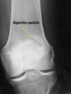
If the various parts of the patella do not join normally as you grow then you can end up with something called a bipartite patella. This is usually asymptomatic but sometimes can require removal of the separated fragment or internal fixation if it is big enough.